Ancient Blood Vessels Discovered in T. Rex Fossil Rewrite Dinosaur Science
Powerful synchrotron X-rays reveal preserved circulatory system in 66-million-year-old Tyrannosaurus rex bone, offering unprecedented insights into dinosaur biology and healing processes.
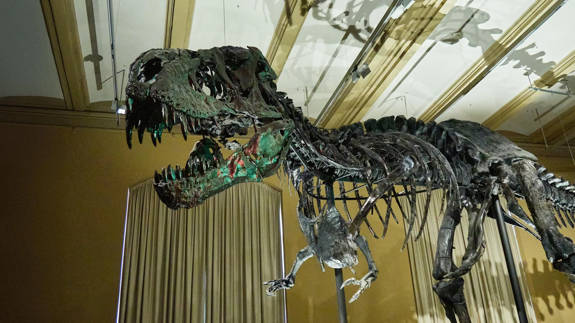
Scientists have discovered an extraordinary network of preserved blood vessels inside a fossilized rib from Scotty, the largest Tyrannosaurus rex ever found, using advanced synchrotron imaging technology that allowed them to peer inside the ancient bone without damaging it. The discovery, published in Scientific Reports, represents a breakthrough in paleontology that could revolutionize understanding of dinosaur biology and the healing processes of these massive predators. The preserved vessels appear to be part of the healing response to a large fracture that had only partially healed when Scotty died 66 million years ago.
The research began when an undergraduate physics student at the University of Regina noticed unusual structures while using particle accelerator technology to examine fossils. Nearly six years later, the same researcher, now pursuing a PhD, has continued applying physics-based methods to improve fossil analysis techniques. Scotty, housed at the Royal Saskatchewan Museum in Canada, proved to be an ideal subject for this groundbreaking research due to evidence suggesting the massive dinosaur lived a difficult life marked by numerous injuries, possibly from combat with other dinosaurs or disease.
The preservation of soft tissue structures like blood vessels inside dinosaur bones presents two major technical challenges that the research team overcame through innovative approaches. First, researchers needed to examine the interior of the fossil without damaging the irreplaceable specimen. Second, fossilized bones are extremely dense because minerals have replaced the original organic material over millions of years, making them difficult to penetrate with standard imaging techniques. The team initially considered conventional CT scanning but found that standard medical scanners cannot penetrate the dense structure of large fossils.
Instead, the researchers turned to synchrotron light, a powerful form of high-intensity X-rays produced at specialized particle accelerator facilities. This cutting-edge technique allowed them to visualize tiny internal features such as blood vessels with remarkable clarity while also analyzing the chemical composition of the preserved structures. The vessels had been preserved as iron-rich mineralized structures that formed a dense network throughout the healing bone tissue, providing a three-dimensional map of the ancient circulatory system.
The implications of this discovery extend far beyond a single specimen, potentially opening new avenues for understanding dinosaur physiology, behavior, and the biological processes that governed their lives. When bones are damaged, the body increases blood vessel activity in affected areas to support healing, and the structures observed in Scotty's rib appear to be part of that ancient healing process. This preserved evidence of dinosaur medical responses provides unprecedented insights into how these massive creatures recovered from injuries and adapted to the challenges of their environment millions of years ago.