Ancient Blood Vessels Discovered in T. Rex Fossil Reveal Dinosaur Healing Secrets
Powerful synchrotron X-rays uncover preserved blood vessels in 66-million-year-old Tyrannosaurus rex rib, providing new insights into dinosaur biology and recovery.
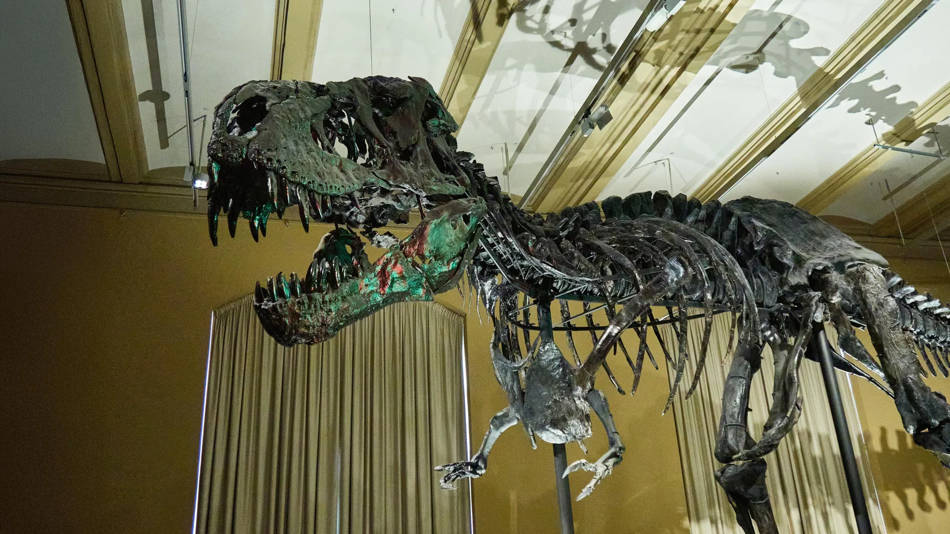
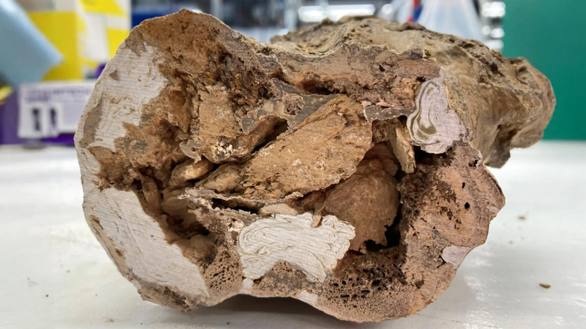
Scientists have discovered preserved blood vessels inside a Tyrannosaurus rex fossil using powerful synchrotron X-rays from particle accelerators, revealing intricate details about how these massive predators healed from injuries 66 million years ago. The preserved vessels were found in a rib from an extraordinary specimen known as Scotty, housed at the Royal Saskatchewan Museum in Canada and recognized as the largest T. rex ever discovered. The rib showed evidence of a large fracture that had only partially healed, and the researchers were able to reconstruct a dense network of mineralized vessels using advanced 3D modeling techniques.
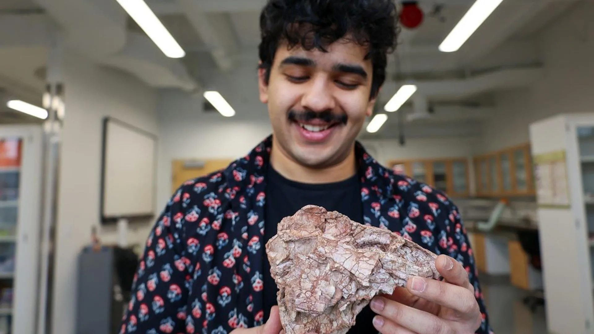
The discovery began when an undergraduate physics student at the University of Regina joined a research group using particle accelerators to study fossils. During advanced 3D imaging of the T. rex bone, structures that appeared to be blood vessels were identified, leading to nearly six years of continued research applying physics-based methods to improve fossil analysis. The breakthrough demonstrates how interdisciplinary approaches combining physics and paleontology can reveal hidden biological processes that occurred millions of years ago.
Scotty lived a particularly difficult life around 66 million years ago, with many bones showing signs of injury possibly from combat with another dinosaur or from disease. When bones are damaged, the body increases blood vessel activity in the affected area to support healing, and the structures observed in Scotty's rib appear to be part of that ancient healing process. The dense network of mineralized vessels provides unprecedented insight into how these massive predators' bodies responded to trauma and attempted to repair themselves.
Studying the internal structure of fossil bones presents significant technical challenges, requiring methods that can penetrate extremely dense fossilized material without causing damage. Standard medical CT scanners cannot penetrate the dense structure of large fossils, so the team used synchrotron light, a powerful form of high-intensity X-rays produced at specialized particle accelerator facilities. This technique allowed researchers to visualize tiny internal features such as blood vessels with remarkable clarity while also analyzing their chemical composition.
The vessels had been preserved as iron-rich mineralized structures, providing chemical evidence of their biological origin and function in the healing process. This discovery opens new possibilities for understanding dinosaur physiology, injury recovery, and the preservation of soft tissues in ancient fossils. The research demonstrates that even after 66 million years, fossils can still yield surprising insights into the daily lives and biological processes of extinct animals, challenging the traditional view that only hard tissues like bones and teeth can provide meaningful information about dinosaur biology.