Flu Virus Dissolves Tiny Droplets in the Cell’s Nucleus to Hijack It, Scientists Discover
Researchers found that influenza A deliberately breaks down structures called paraspeckles inside the nucleus, freeing proteins the virus appears to exploit to replicate — a discovery that could point to new antiviral drugs.

Scientists have uncovered a striking new trick that the influenza A virus uses to commandeer the cells it infects: it dissolves tiny droplet-like structures deep inside the cell's nucleus, releasing a cache of proteins that the virus appears to turn to its own advantage.
The work, published July 21 in the journal Nature Microbiology, was led by researchers at the European Molecular Biology Laboratory (EMBL) in Hamburg, working with colleagues at the Leibniz Research Institute for Molecular Pharmacology in Berlin and the Charité's Institute of Virology. Using a combination of cutting-edge techniques, the team mapped in unprecedented detail how the virus rearranges the inner machinery of a human cell.
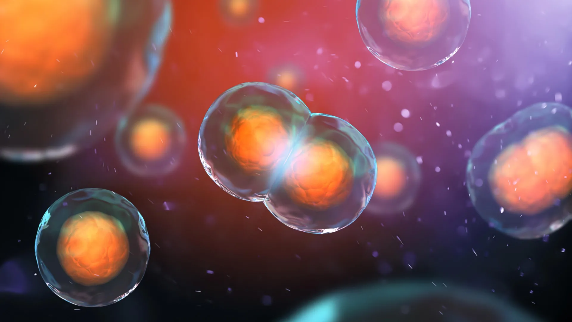
At the center of the discovery are paraspeckles — small, membraneless compartments in the nucleus that form like droplets and help regulate the activity of genes and RNA-binding proteins. The researchers found that infection with influenza A causes these paraspeckles to fall apart. Crucially, the effect was not a random casualty of infection but a consistent, repeatable event. "Watching these tiny organelles in the nucleus dissolve, consistently across every cell line and every flu strain we tested, told us this isn't a side effect," said Iuliia Kotova, the study's first author, who conducted the work at EMBL Hamburg and is now at ETH Zurich.
When the paraspeckles dissolve, they release RNA-binding proteins that had been sequestered inside them. The team's evidence suggests the virus may harness those freed proteins to help copy its own genetic material, while the breakdown may simultaneously weaken the cell's antiviral defenses — a double benefit for the invader. In effect, the virus appears to dismantle part of the cell's organizational system and repurpose the pieces.
To capture these interactions as they happened, the scientists used cross-linking mass spectrometry inside intact cells, a method that essentially freezes proteins in place while they are touching, and combined it with structural modeling from the artificial-intelligence tool AlphaFold. That allowed them to build a molecular map of which proteins were interacting during an active infection, revealing the choreography behind the takeover.
The researchers, including group leaders Jan Kosinski at EMBL Hamburg and Boris Bogdanow at the Charité, say the detailed map could expose new vulnerabilities to target with drugs and inform the design of more effective vaccines and antiviral treatments. That matters most for dangerous pandemic-potential strains such as H5N1 bird flu, which has alarmed public-health officials as it spreads among animals. By understanding exactly how the flu bends a cell to its will, scientists hope to find fresh ways to throw a wrench into the process.