Physicists Imaged an Electron's Wavefunction in 3D Using a Laser That Fits on a Table
The measurement used to require weeks at a synchrotron. A Göttingen team rebuilt the reconstruction algorithm from scratch and did it in a university lab, resolving detail finer than the gap between two carbon atoms.
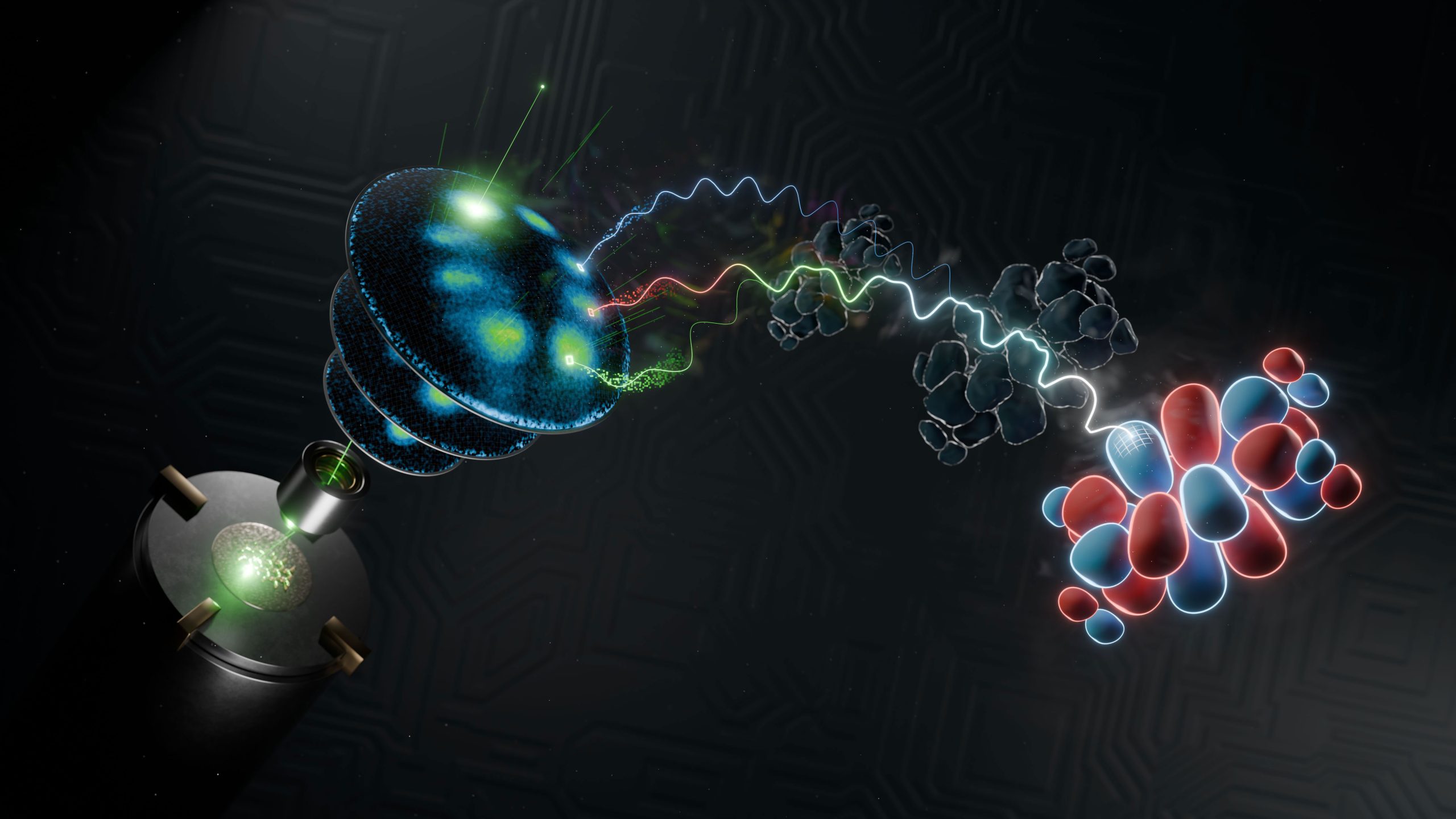
The wavefunction is the central object in quantum mechanics and the one you are not supposed to be able to look at. It encodes everything knowable about a particle, but it is not itself a measurable quantity — what experiments return are probabilities derived from it. Physicists at the University of Göttingen have now produced a three-dimensional image of one, for the electron orbitals of a single organic molecule, using equipment that sits on a laboratory bench.
"The wavefunction is a fundamental quantity in quantum mechanics, yet it cannot be directly observed or measured," said Professor Stefan Mathias, who led the work. The results were published in Nature Communications.
The technique is photoemission orbital tomography. Short pulses of extreme ultraviolet light knock electrons out of a molecule; detectors record the direction and energy of every escaping electron, building up a map in momentum space. That map is mathematically related to the shape of the orbital the electron came from, and with enough angles the orbital can be reconstructed — the same logic as a medical CT scan, run on the quantum state of a molecule instead of a body.
Doing it in two dimensions has been possible for over a decade. Going to three has meant booking time at a synchrotron — a building-sized accelerator ring — and collecting data for long stretches, because the reconstruction demanded an enormous number of measurements from many orientations. The scarcity of beam time is why the method never spread far beyond a handful of groups.
The Göttingen team removed both bottlenecks at once. They paired a lab-based soft X-ray source producing ultrashort pulses with a reconstruction algorithm they rewrote from the ground up. "We introduce two powerful new concepts," said Dr. Matthijs Jansen, a co-leader of the study. "Reliable 3D images can now be obtained using much less experimental data." The demonstration target was PTCDA, a flat organic molecule familiar to surface physicists and used industrially as a red dye. The reconstruction resolves features smaller than the spacing between neighboring carbon atoms inside it.
The payoff the group is chasing is motion. Because the light source delivers femtosecond pulses — quadrillionths of a second — the same setup can in principle take a series of snapshots as a molecule absorbs light and its electrons rearrange. "This technique might mean that stroboscopic videography becomes a reality," said Dr. Wiebke Bennecke, allowing researchers to watch not just the shape of a wavefunction but its evolution.
That matters for anything that depends on what electrons do in the first instants after a photon arrives: organic solar cells, photocatalysts, molecular electronics, the initial steps of photosynthesis. Those processes are currently inferred from spectroscopic signatures and modeled computationally. Filming the orbitals directly would replace the inference with a measurement, and the fact that it now takes a table rather than an accelerator ring is what determines how many laboratories get to try.


